


First impressions of MCC’s revolutionary scanner
CCM - Fitte
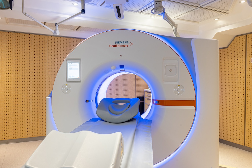
The scanner was inaugurated on December 16, in the presence of Prince Albert II. Dr. Filippo Civaia and Dr. Philippe Rossi present the revolutionary device and share their first impressions.
The project had been in development for twenty years. Today, Monaco’s cardiothoracic centre boasts one of the first photon counting scanners in the world to be entirely dedicated to cardiovascular pathologies.
“The objective is to provide better care for our patients,” explains Dr. Civaia, one of the centre’s cardiologists. “This scanner is the culmination of a pioneering joint project between Monaco, Germany and the United States. The aim was to use photon counting as a new means of capturing images.”
The doctor uses an image (!) to explain the complex process. “When you use a standard scanner, it’s like using a watering can without its rose. When you water your plants, some of the water will be poured where you need it, but some goes elsewhere. With the new scanner, you add the rose to the watering can and you can pour the water with a finer spray. You can then break down the different lines of spray and analyse them better.”


Avoiding invasive treatments
The objective is therefore to see better, but above all to understand the images that appear on the screen better. “We can get a better handle on the plaque [made up of lipids, fibrous tissue and calcium] in the coronary arteries and judge whether it is stable or unstable, because all cardiology issues are plaque-related,” adds Dr Civaia. “And with this new technique, you can look more closely at the components of the plaque and identify those that could cause an acute cardiac episode.”
For patients, the benefits are significant: with much more accurate images, cardiologists are able to better define treatment. This helps to increase efficiency. The contrast media used to perform the scan and the examination time remain the same.
With more accurate diagnoses, some patients will be able to avoid the use of other invasive complementary examinations, such as coronary angiography which involves puncturing an arterial vessel, hospitalisation and, potentially, complications.
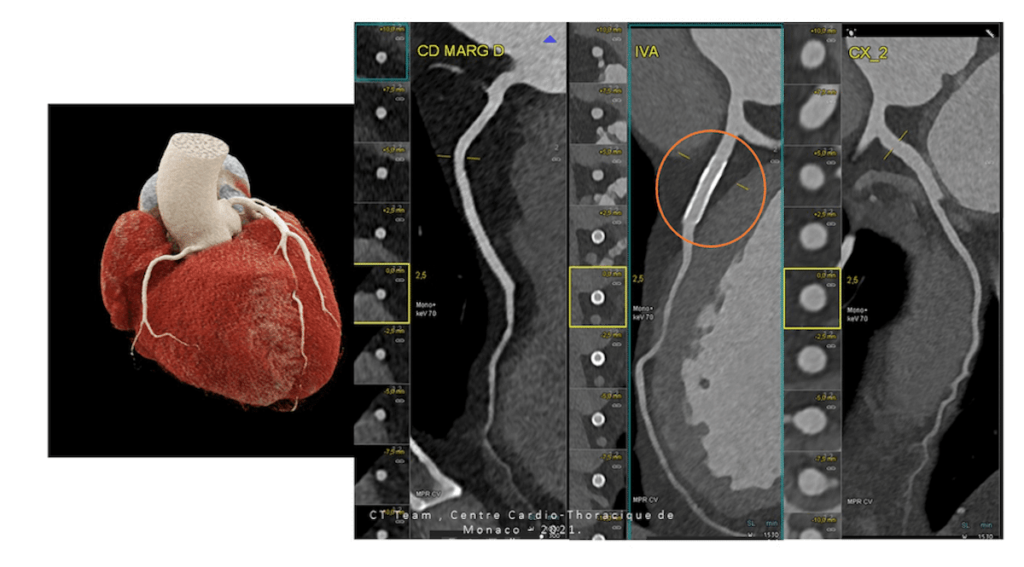
Excellent patency and functionality of a coronary stent 
Patient with advanced coronary artery disease, as evidenced by the presence of several plaques: excellent correlation between CT scan and traditional coronary angiography.
It is in everyone’s interests for examinations to be as low-radiation as possible
Reduce the amount of radiation
At the same time, Dr. Rossi, cardiologist, mentions a considerable advantage: “We also have a reduction in radiation used on patients, with an irradiation rate that is reduced by a factor of 10 compared to the first cardiac scanners. When we undergo a radiology examination using X-rays, we never lose that dose of X-rays and if these build up to over a certain level, we risk developing radiation-induced pathologies, in particular cancer. It is in everyone’s interests to make examinations as low-radiation as possible”.
More than 1,000 examinations have been carried out at the centre since the commissioning of the new scanner in September. Despite a lower radiation level than standard scanners, doctors Civaia and Rossi point out that there is still radiation involved, so the examination must be prescribed by a doctor. Its use as a preventive measure is therefore not envisaged for the moment: it is a diagnostic tool.
Other institutions around the world are also equipped with these revolutionary scanners, but in other fields such as neurology or pneumology.